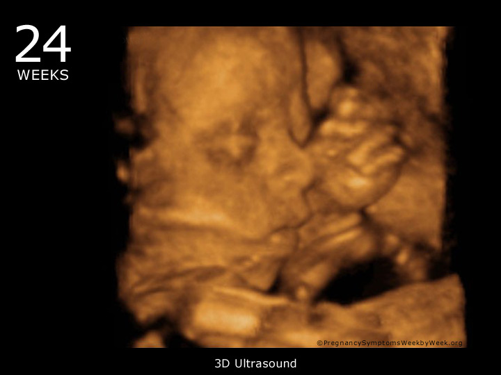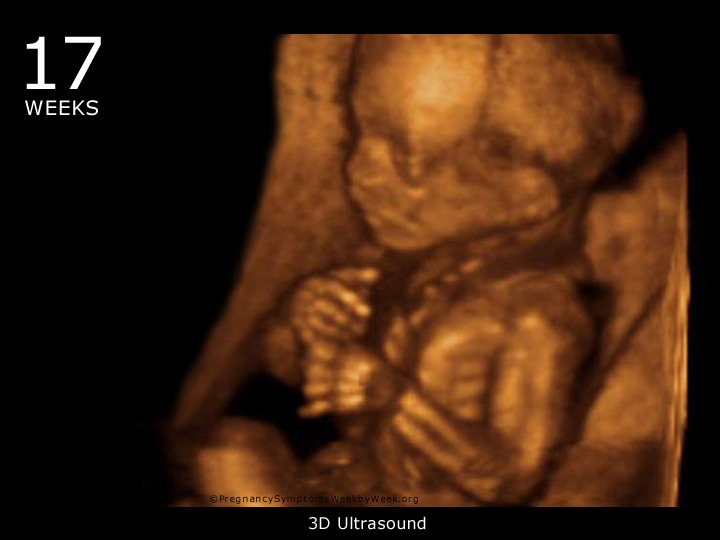
Department of Labor.Īmerican College of Obstetricians and Gynecologists. Vitamin D: screening and supplementation during pregnancy. Vitamin D: Fact Sheet for Consumers.Īmerican College of Obstetricians and Gynecologists. National Institutes of Health Office of Dietary Supplements. Benefits of docosahexaenoic acid, folic acid, vitamin D and iodine on foetal and infant brain development and function following maternal supplementation during pregnancy and lactation. 5600 W Maple Rd B-206 West Bloomfield, MI 48322. 3D Ultrasound 4D 5D HD HDlive Ultrasound & Early Gender. All of these images were taken here at SonoSmile which does amazing 3D ultrasound in Ocala. Fetal Development Milestones: At 27 weeks, your baby-to-be is at the end of the second. Are There Benefits to Spending Time Outdoors?. Find out if you’re having a girl or boy 15 Min 2D Ultrasound. This page shows typical 3D ultrasound images from 11 to 36 weeks. Fetal Size: Length, 9 2/3 inches (crown to rump) total length about 15 1/4 inches weight, 2 pounds. Division of Cancer Prevention and Control. doi:10.1038/nature11823Ĭenters for Disease Control and Prevention. A direct and melanopsin-dependent fetal light response regulates mouse eye development. Prospective evaluation of nighttime hot flashes during pregnancy and postpartum. It results in a baby born without signs of life. Stillbirth is defined as fetal death after 20 or 28 weeks of pregnancy, depending on the source. The underlying cause in about half of cases involves chromosomal abnormalities. I just want my baby to be OK, I repeated over and over again on a Thursday morning last April. Thurston RC, Luther JF, Wisniewski SR, Eng H, Wisner KL. About 80 of miscarriages occur in the first 12 weeks of pregnancy. My 28-Week Ultrasound Confirmed My Worst Nightmare. Fetal response to sound and light: Possible fetal education. Fetal Development.Īmerican College of Obstetrics and Gynecologists. Earlier Ultrasounds (24 to 28 weeks) allow the expectant mother to see more whole body images of her baby. Photos of belly growth at 17 weeks pregnancy.

RF 2C7J445 Pregnant young woman with pregnancy week number next to her belly. RM BA0738 This stock medical exhibit depicts a twenty four week old fetus. The World Health Organization Fetal Growth Charts: A multinational longitudinal study of ultrasound biometric measurements and estimated fetal weight. The best time is between 24-32 weeks gestation. RM E82FKD Foetus' face, Coloured 3-D ultrasound scan of a foetus, Gestational age : 24 weeks.


Reference values for valve circumferences and ventricular wall thicknesses of fetal and neonatal hearts.


 0 kommentar(er)
